24 May A Snapshot With a Higher Purpose: High-Resolution Images in Ophthalmology
The old saying “a picture is worth a thousand words” couldn’t be more true: aside from capturing the beauty of special occasions, images are also used in the ophthalmology field to document a specific eye condition’s progress by freezing a moment in time.
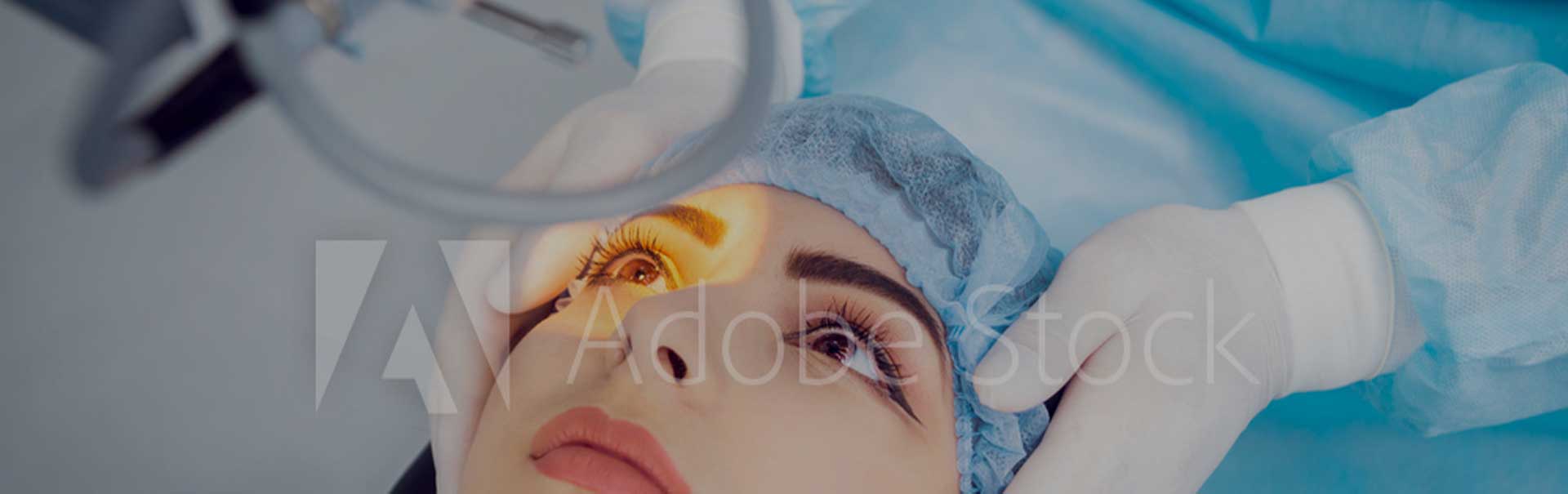
We use Topcon’s specialized slit lamp camera to document such ocular conditions as pterygium, corneal ulcer, herpes keratitis, and tumor. After determining that an image would be beneficial for a patient, the baseline image is captured within a split second – there is no discomfort or dilation necessary.
Benign growths (referred to as iris nevi) can be found in the colored part of the eye. These growths, which rarely develop into malignancies, are kept in check via these specialized images and incremental follow-ups (usually 6 months to a year).
In addition, these high-resolution images are used as an educational tool so patients can see the severity of conditions such as Blepharitis, where persistent inflammation of the eyelids sometimes presents irritation, itching, and red eyes. On follow-up visits, comparing pre- and post- treatment images shows progress that is not visible to the naked eye. Patients can appreciate first-hand the benefits of adhering to the recommended warm compress, careful daily cleansing of lashes, artificial tears, and/or antibiotic regiment outlined by their physician.
Essentially, the team at Del Negro & Senft Eye Associates can scrutinize every minute change from one exam to the next with a high-resolution image. In the past, doctors tracked these specific conditions/variations by writing down and measuring the findings in the patient’s chart. But now, our team can simply compare images in real time to document and track progression with incredible accuracy.
Sorry, the comment form is closed at this time.