Advanced Technology

Our Services

Thanks to advances in technology, even surgeries that just a few years ago required hospitalization can now be handled safely and with greater comfort and convenience in a surgery center.

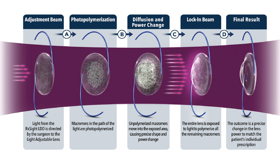
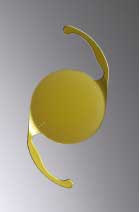
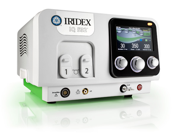
Doctors Ralph Del Negro, Carl Senft, and Marina Glatman lead the way in ophthalmology technology innovation, with Laser Vision Correction/PRK and Laser Cataract Surgery with advanced technology lens options. Our doctors perform cataract and glaucoma procedures and sophisticated corneal surgery at Seashore Surgical Institute in Brick.*

They focused on your needs when planning and equipping this accredited, full-service facility that makes it possible for them to deliver better results and increase the comfort and satisfaction of their patients. From registration to discharge, a streamlined administrative process makes your eye surgery a more convenient and less stressful experience. When appropriate, Drs. Del Negro, Senft, and Glatman also perform procedures at Amdur Ambulatory Care Center, a part of Hackensack Meridian Health Jersey Shore University Medical Center in Neptune, NJ.

*Drs. Del Negro and Senft have an ownership interest in Seashore Surgical Institute.

LenSx® Femtosecond Laser for Laser Cataract Surgery

Advanced Technology Lens Options

Allegretto (OptiLasik) utilized for Laser Vision Correction/PRK

IRIDEX MicroPulse® Laser for treatment of Open Angle Glaucoma & Retinal Diseases

Del Negro & Senft Eye Associates makes use of the following technology:

Click to learn more.

ARGOS

The ARGOS® swept-source optical coherence tomography (SS-OCT) biometer gathers the required diagnostic measurements for Cataract or Laser Vision Correction surgery by capturing a high-resolution image of each patient’s unique eye. The ARGOS is a non-invasive, non-contact biometer that is used to acquire ocular measurements as well as perform calculations to determine the appropriate intraocular lens (IOL) power and type for implantation during intraocular lens placement. ARGOS measures the following 9 parameters: Axial Length, Corneal Thickness, Anterior Chamber Depth, Lens Thickness, K-values (Radii of flattest and steepest meridians), Astigmatism, White-To-White (corneal diameter) , and Pupil Size.

PENTACAM

A rotating Scheimpflug camera captures multiple, high resolution images providing valuable information on Refractive, Cataract, Glaucoma, and Corneal findings. Within a mere second the Pentacam® supplies our doctors with precise diagnostic data on the entire anterior eye segment. For those diagnosed with a cataract, the degree of cataract is made visible by the light scattering properties of the eye lens. Measurement of the anterior and posterior corneal surfaces supplies the true total refractive power of the cornea. The data on the posterior surface provides information needed for early keratoconus detection, a test Dr. Senft orders to determine if a patient is a candidate for Laser Vision Correction. The rotating scan supplies more data points in the center of the cornea. For added accuracy, a supplementary pupil camera corrects for eye movement during the examination. Pentacam.com

FLUORESCEIN ANGIOGRAPHY

Dr. Glatman utilizes fluorescein angiography to aid in the diagnosis and management of various conditions that may occur in the retina. Pictures taken after a dye is injected help pinpoint signs of retinal circulation problems, swelling, leakage, or abnormal blood vessel formation, which helps guide treatment.

View Video

OPTOS-Retinal Photography

Annual eye exams are vital to maintaining your vision and overall health. Our doctors offer the optomap® as an important part of this screening process. The first time a new patient is seen in the office our doctors dilate as normal, establishing a baseline. Following this initial dilated exam, a welcome benefit to our patients is that that a routine exam can be performed without dilation, if there are no additional findings to suggest otherwise. The optomap produces an image that is unique and provides Drs. Del Negro, Senft, Glatman, Shah, and Carniglia with a high-resolution 200° image in order to ascertain the health of your retina. This is much wider than a traditional 45° image.

Many eye problems can develop without you knowing, in fact, you may not even notice any change in your sight – fortunately, diseases or damage such as macular degeneration, glaucoma, retinal tears or detachments, and other health problems such as diabetes and high blood pressure can be seen with a thorough exam of the retina.

The inclusion of optomap as part of a comprehensive eye exam provides:

  • A scan to show a healthy eye or detect disease.
  • A view of the retina, giving your doctor a more comprehensive view than they can get by other means.
  • The opportunity for you to view and discuss the optomap image of your eye with your doctor at the time of your exam.
  • A permanent record for your file, which allows our doctors to view your images with you each year to look for changes.

The optomap is fast, easy, and comfortable for anyone. The entire image process is simple–you just look into the device one eye at a time.

Spectralis/OCT

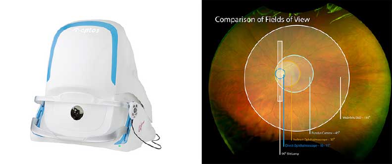
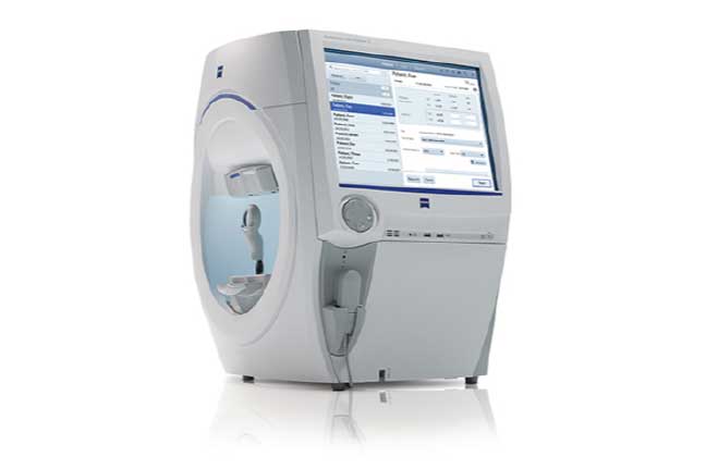
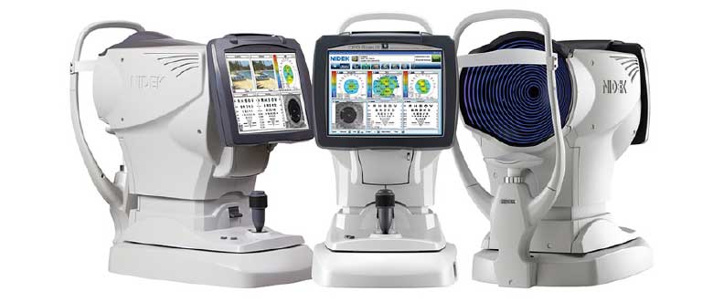
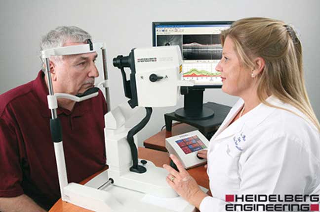
Del Negro & Senft Eye Associates is making a difference in the community fight against sight-stealing eye diseases such as glaucoma and macular degeneration (AMD). It is well known that the key to preventing vision loss is early detection. With the new addition of this 3-D imaging technology, our doctors are now better able to detect and manage glaucoma and retinal disease such as macular degeneration and diabetic retinopathy.

This new system, known as SPECTRALIS, uses an advanced eye tracking system that produces 3-D images of the eye, allowing our doctors to more easily identify change over time. This helps reveal eye disease which may not yet be noticeable by the patient or detectable with a traditional visual exam. The SPECTRALIS not only aids in the detection of such diseases as glaucoma and macular degeneration, it also provides essential information for monitoring them in follow-up examinations.

The SPECTRALIS exam is fast, safe and usually does not require dilating the eye. While a patient comfortably sits in a stationary position, the eye is scanned in a few seconds.

Visual Field

The visual field test is the standard of care in glaucoma diagnosis and management. It provides proven diagnostic performance in detecting early visual field loss. It is also helpful in neurologic diagnosis, retinal diagnosis, as well as diagnosing other visual problems.

Humphrey® Field Analyzer 3, Image courtesy of ZEISS

OPD Scan (Optical Coherence Tomography)

At Del Negro & Senft Eye Associates, our team uses the innovative OPD III Wavefront system. This remarkable instrument offers the most advanced digital technology available to help us better evaluate your unique needs and provide you with the best possible vision. Far more than just a refractive examination, this instrument provides a comprehensive understanding of your complete visual system and optical pathway. Using information from the OPD III Wavefront system, our team can make better recommendations to help you see better in all types of settings.

How Is Your Current Vision?

  • Trouble with excessive glare and less than optimal vision when driving at night?
  • Feel like you just aren’t seeing your best with your latest pair of glasses?
  • Think cataracts are impacting your vision?

OPD testing can provide insights that contribute to developing a proper treatment plan. Our team is thrilled to offer the best technology and care for our patients. Our job is to provide you with your best possible vision.

The OPD-Scan III performs at least five important clinical functions:

  • Automated refraction
  • Automated keratometry
  • Corneal topography
  • Pupillometry
  • Wavefront aberrometry

Refraction-Marco COS Unit

This state-of-the-art system has the capability of allowing our doctors to make a comparison between your current refractive error, your old prescription, and your new prescription. Vision diagnostic results are faster, yet more accurate than ever. The system combines smart features such as an electronic refractor, auto refractor/keratometer, chart projector, and auto lensmeter. The ergonomic design ensures that patients are comfortable while the technician is performing a thorough work-up. The end result is a precise diagnosis of vision exceeding expectations for consistency and accuracy.

Photo-Slit Lamp

The slit lamp camera is used to document disease states of the front of the eye, patient education, and monitoring.

Light Delivery Device (LDD)

LDD Treatment

Lens Technology & LDD Treatment

Request an Appointment

Our goal is to contact you by phone within 24 hours of submission. If you find yourself in an EMERGENCY situation, please CALL 732-774-5566 instead.

Skip to content

LenSx® Femtosecond Laser for Laser Cataract Surgery


No matter what you value most in life – working, reading, golfing, watching your children or grandchildren, or just taking in the scenery – the level of enjoyment you get out of those activities is very dependent on your sight. If your vision is clouded by cataracts, you may be limited in enjoying the things that are most important to you. Cataract surgery can significantly improve your quality of life.

When you choose cataract surgery with Drs. Del Negro, Senft, and Glatman, we make sure you have the best and most advanced options available to you. That’s why we are excited to be the first eye surgeons in the area to offer Laser Cataract Surgery to provide our patients the opportunity to achieve their best possible vision.

WHAT IS THE LENSX® LASER?

The LenSx® laser is a femtosecond laser that gives Drs. Del Negro, Senft, and Glatman computer controlled precision, allowing more accurate performance of some of the key steps of cataract surgery. Using a variety of highly advanced technologies, the LenSx captures precise, high resolution images of your eye. To further enhance accuracy, a patient interface connects your eye to the image-guided unit so that the LenSx laser computer and the surgeon commanding it have precise, real time images of your eye at all times during the procedure. These images allow the surgeon to accurately guide the laser in performing cataract surgery with a level of precision not previously possible.

LenSx® IS CUSTOMIZED TO YOUR EYES

All human eyes may share the same basic anatomic structure, but every eye is unique in terms of size, depth and curvature of the cornea, size of the lens, and length of the eye. These parameters are carefully measured and mapped prior to surgery. The LenSx laser uses optical coherence tomography to capture the precise, high resolution images of your eye. This, along with the information obtained prior to surgery, allows Drs. Del Negro, Senft and Glatman to plan and perform surgery specific to each eye.

With traditional cataract surgery, the incision is made with a blade and access to the cataract is obtained using a needle. Not only does the LenSx laser create extremely precise cataract incisions and break up the cloudy lens for easier removal, it also provides the surgeons with the ability to correct/improve astigmatism with more accuracy and precision than ever before.

Drs. Del Negro, Senft, and Glatman’s expert micro-surgical technique, along with LenSx’s gentle, accurate, and precise features strengthen their reputation for providing premier cataract surgery in Monmouth County.

THE ADVANTAGES ARE CLEAR

  • The femtosecond laser makes more precise incisions when compared to the traditional method.
  • The LenSx laser technology gives Drs. Del Negro, Senft, and Glatman the ability to reduce astigmatism with greater accuracy. This allows for better performance of the advanced technology lenses and, ultimately, provides the best possible visual outcome.
  • Laser precision creates a uniform, circular opening in the capsular bag that is perfectly centered; this is critical for effective lens position to ensure your best visual outcome.


Advanced Technology Lens Options


If I'm a candidate, what lens options will my surgeon recommend?


Introducing the RxSight® Light Adjustable Lens

Treats: Presbyopia, astigmatism, excellent visibility in all lighting conditions.

RxSight® Light Adjustable Lens

The RxSight® Light Adjustable Lens is the first and only intraocular lens (IOL) that can be customized after cataract surgery. The Light Adjustable Lens is made of a special photosensitive material that can be adjusted in response to ultraviolet (UV) light. This optimization is completed by Dr. Del Negro in the weeks following lens implantation through a series of non-invasive light treatments that take only a few minutes each.

You will have the unique ability to adjust and preview your vision until it meets your personal desires and lifestyle requirements. The Light Adjustable Lens delivers superior vision outcomes that non-adjustable IOLs cannot match.

You may need 3 to 5 total light treatments over a period of 4 to 5 weeks to reach your vision goals.

UV-Blocking Glasses

During the period immediately following surgery until 24 hours after your last light treatment is completed you will be required to wear special UV-blocking glasses during all waking hours. Wearing ONLY the glasses we supply will prevent exposure to both INDOOR and OUTDOOR sources of UV light that can cause uncontrolled changes to the Light Adjustable Lens.

Detailed instructions will be provided.

Presbyopia Correction at the time of Cataract Surgery

If you are interested in pursuing functional vision for normal, everyday activities a presbyopia correcting IOL may be the right choice for you such as Alcon’s AcrySof® IQ PanOptix®, or Johnson & Johnson’s TECNIS®. A presbyopia correcting intraocular lens (IOL) allows for correction of vision at near, intermediate, and distance and provides a full range of high-quality vision at functional distances. It supplies uninterrupted vision and less dependence on glasses when driving, using your computer, or using your mobile device.

Two Options:

#1

  • Corrects for presbyopia
  • Excellent visibility in all lighting conditions

 
#2

  • Corrects for presbyopia AND astigmatism
  • Excellent visibility in all lighting conditions


CLEAR, COMPLETE VISION


UP CLOSE

Crafting, reading a book or magazine


ARM’S LENGTH

Read a menu, see yourself in the mirror


FAR AWAY

Recognize a familiar face, drive, watch TV, see a show

Your surgeon will carefully evaluate the visual demands of your individual lifestyle before recommending a particular lens. If you have astigmatism, your surgeon may recommend a Toric or presbyopic-correcting lens designed to correct that condition. There are circumstances where another lens option may be more beneficial for a particular patient. All options would be discussed during your exam.

Astigmatism Correction at the time of cataract surgery?

A person who has both a cataract and corneal astigmatism will not regain quality distance vision after cataract surgery unless the astigmatism is also corrected. Alcon’s AcrySof® IQ TORIC or Johnson & Johnson Vision’s TORIC Tecnis® II lens makes it possible to correct astigmatism and cataracts at the same time and provides quality distance vision. Its unique design makes it possible to reduce, or eliminate, corneal astigmatism and significantly improve uncorrected distance vision.

Who should consider having an advanced technology replacement lens implanted?

Candidates for these advanced-technology lenses include patients age 45 or older who desire a full range of vision in each eye with less dependence on reading glasses, bifocals, or progressive lenses as well as individuals with cataracts that impair vision. Patients with health problems, such as diabetes or a chronic infection, should wait until those conditions are under control before scheduling the procedure.

Drs. Del Negro, Senft, or Glatman will perform a careful examination before making any recommendation.

Learn More About Cataract Surgery


Allegretto (OptiLasik) utilized for Laser Vision Correction/PRK


EXPERIENCE CUSTOMIZED VISION

We know laser vision correction is an important decision, so Del Negro & Senft Eye Associates takes great care to determine what’s best for you, our patient. That’s why we offer OptiLASIK™ Laser Vision Correction.

OPTILASIK™ LASER VISION CORRECTION GOES BEYOND TRADITIONAL LASER VISION CORRECTION TREATMENTS

Even though everyone’s eyes are unique, in the past, Laser Vision Correction surgery had to be pretty much the same for everyone. It simply corrected your vision based on your current glasses or contact lens prescription. But OptiLASIK™ Laser Vision Correction is much more advanced than that.

It uses the innovative diagnostic technology of the Allegretto Wave® Eye-Q Laser System to create a detailed “map” of your eyes. By using your map as a guide, Dr. Senft is able to personalize the treatment for your best results, quickly and safely. And because OptiLASIK™ Laser Vision Correction is so precise and accurate, it opens the doors for many people with myopia, hyperopia, and astigmatism who weren’t eligible for laser vision correction in the past.

OPTILASIK™ LASER VISION CORRECTION ASSURANCE AND COMFORT

The OptiLASIK™ Laser Vision Correction method has been used successfully on hundreds of thousands of eyes with exceptional results. Our commitment is to provide you with the ultimate in comfort, safety, and outstanding vision. OptiLASIK™ Laser Vision Correction can help you achieve all of this.

THE OUTCOMES SPEAK FOR THEMSELVES

OptiLASIK™ Laser Vision Correction patients describe their vision as nothing short of life changing. After surgery, most patients can see as well as, or even better than, they did with glasses or contact lenses: 93% of nearsighted patients in the clinical trial submitted for approval of the laser saw 20/20 or better and 83% of farsighted patients saw 20/25 or better. What’s more, patients were so happy with the results that 97% would recommend OptiLASIK™ Laser Vision Correction to their friends.

With its surgical accuracy and ability to address your unique vision needs, OptiLASIK™ Laser Vision Correction could completely change the way you see the world. Call us at 732-774-5566 x239 to find out if it’s right for you.

Learn more about Laser Vision Correction/PRK


IRIDEX MicroPulse® Laser for treatment of Open Angle Glaucoma & Retinal Diseases


Indications- The MicroPulse Laser Trabeculoplasty (MLT) surgeries are used for patients with open angle glaucoma. The laser is utilizes to treat the drainage system of the eye known as the trabecular meshwork. Treating this area of the natural internal draining system is designed to improve the outflow of fluid from the eye.

This type of laser surgery will be effective in some patients, but not others. Your response is determined by the type of glaucoma you have and the specific structures found in your drainage system. Your doctor cannot predict how well the laser will work before the laser surgery.

Procedure- The laser machine is similar to the examination microscope that the Ophthalmologist uses at each visit to look into your eyes. The laser itself makes little noise and flashes a light about as bright as the flash on a camera. Nearly all patients find the procedure comfortable and pain free. The procedure generally takes 10 – 20 minutes.

Medications- You may need to use drops both before and after the laser treatment. As the pressure in the eye may temporarily go up after laser treatment, you will likely need to have your pressure measured after the laser surgery. If the pressure does elevate, you may need additional medicines to lower the pressure, which will be administered in the office. Rarely, the pressure in the eye could elevate to a level that may require surgery in the operating room to relieve the glaucoma. You need to use drops after the laser to help the eye heal correctly. In most cases you will be asked to continue your other glaucoma medications after the laser surgery. The doctor will notify you if there are exceptions to continuing your medications.

Risks and complications- Glaucoma laser surgery may be followed by complications. Most patients notice some blurring of their vision after laser surgery. This generally clears within a few hours. The chance of your vision being permanently affected by this laser is very, very small. Although rare and unusual, there may be bleeding within the eye, inflammation, cataract and increase in the pressure in the eye requiring different and more extensive treatment. It will take several weeks to determine how much of your eye pressure will be lowered with this treatment. You may require additional laser surgery to lower the pressure.

Learn More About Glaucoma


The IRIDEX MicroPulse® Laser is also indicated for the treatment of Retinal Diseases.


FOCAL LASER

Focal Laser is used by Dr. Glatman to treat Diabetic Macular Edema. During the treatment, a laser beam is used to seal the leaking blood vessels near the macula.

View Video


PANRETINAL LASER

Panretinal Laser is used by Dr. Glatman to treat Proliferative Diabetic Retinopathy. During the treatment, a laser beam is used to treat the peripheral retina leading to cessation of growth of abnormal blood vessels. More than one treatment may be needed depending on the severity of your diagnosis. Your individualized treatment plan would be discussed during your evaluation with Dr. Glatman.

View Video


RETINAL RETINOPLEXY

Dr. Glatman uses laser pulses to seal the retina around a retinal tear or hole, which then forms scar tissue within two weeks. The benefit of this in-office laser treatment is to decrease the chance of developing a retinal detachment due to a retinal tear.

View Video